
Translated material
Cell from Cell
 1
1 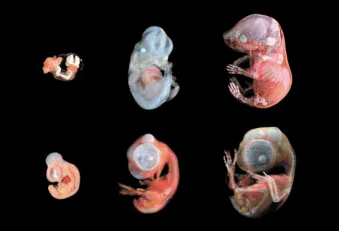 2
2 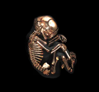 3
3 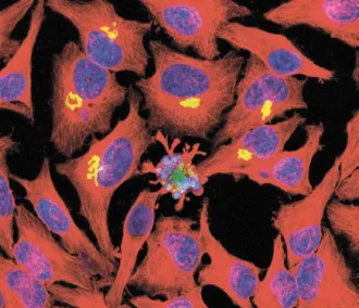 4
4 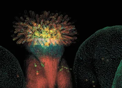 5
5 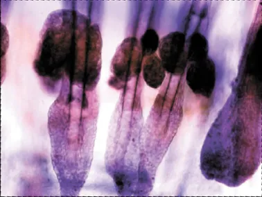 6
6  8
8 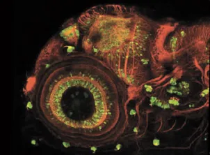 9
9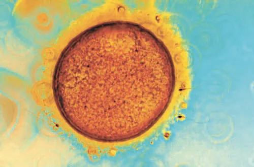 10
10 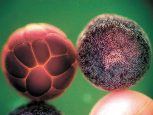 11
11 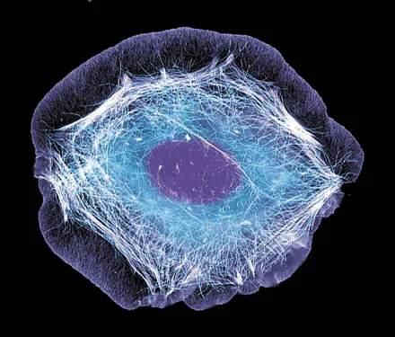 12
12Magazine Cell - This Not Only first -class Scientific publication by molecular And cellular biology And embryology , not only source scientific news For scientific journalists , but And Great popularizer science . On the site publications There is a whole Substed Cell Show [1], on which amazing with German are represented cells , viruses And embryos .
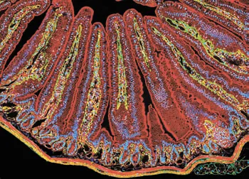
- Mouse intestinal cells.
- A picture taken from mice developed in Kaltekh (there is such a CALTECH MRI Atlas of Mouse Development). This is how the mouse embryos develop (above) and quail (below).
- There are only 206 bones in the body of an adult - and after all, the fetus has more than 300 of their embryonic development at 9 months. This is especially characteristic of the bones of the skull - after all, the brain further grows. Here is a picture of the embryo skeleton in the third trimester of development obtained using computed tomography.
- The line of "immortal" (capable of sharing an infinite number of times) HELA cells obtained from a cancerous tumor.
- Rhizhovida Talya. Fragment of a flower removed with a 20-fold increase using a confocal microscope. Red “shines” the cell membrane, painted with Lipophilic paint FM4-64FX, blue-DNA (dye HoECHST 33342).
- The total drug of the mouse tail epidermis. RNA localization is indicated by purple.
- Drosophila epithelium cancer. And flies have tumors!
- And this is already an anti -cancer drug. Crystals of Mitoxantron in a polarized light.
- A picture of neurons of the eye, ear, brain and side line of the 4-day fry Danio Roerio.
- Failure. This picture, taken using an optical microscope, shows an unsuccessful attempt by IVF. Spermatozoa could not break into the egg.
- Svate frog embryos ( Xenopus Laevis ) at the early stage of embryogenesis. It was removed using an optical microscope and processed with special software. In this picture, the “work” of the SOX3 transcription factor is visible. Those sections of the embryo where this SOX3 expresses will become ectodermal.
- A man of human keratinocyte.
Prepared Alexey Paevsky