
Translated material
Operate before birth
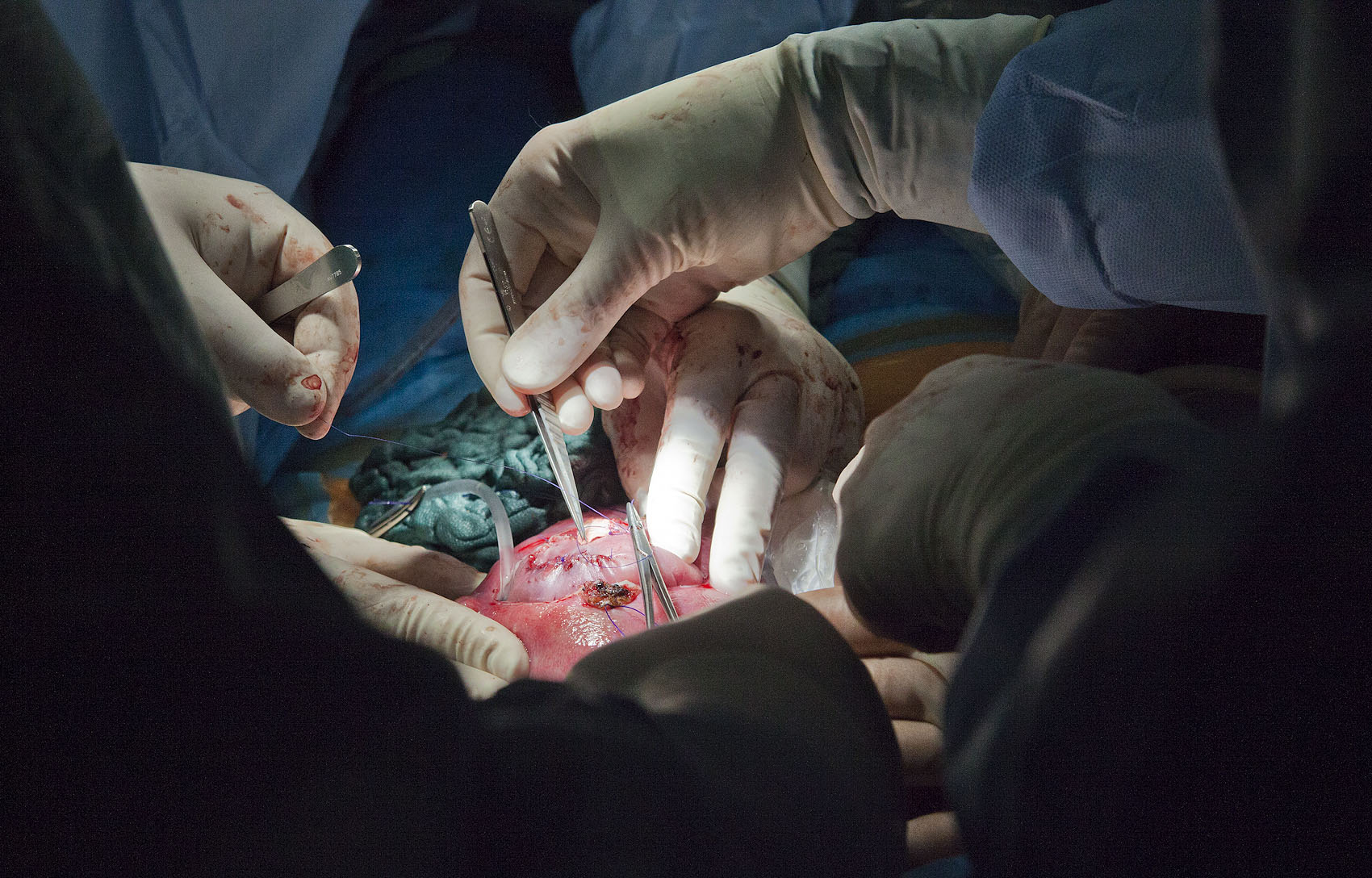
Many pregnant women do not know why the doctor prescribes folic acid to them all. A defect in the formation of the fetal nervous tube, Spina Bifida, Myelomeningocele, D's hernia of the spinal cord-every year and a half or two thousand children patients with this disease are born in Russia. Everyone has problems with the bladder, every second one does not feel the legs, every fifth is paralyzed and has a huge head. Very often, parents refuse children with Spina Bifida. And yes - Spina Bifida can be successfully treated
Help the back of Bifida helpIndications for the operation
In the Operating Zurich University hospital - fifteen people, counting students and assistants. The anesthesiologist is more likely to be like a spaceship control panel. This is a difficult task: to anesthetize the three: a pregnant woman, her uterus, so that she does not come into a tone when they cut, and the child inside, or rather, the fruit with the size from the kitten. Now the woman, her uterus and her unborn child are sleeping. Four inclined above them-a gynecologist surgeon, two neurosurgeons and an operating room. What they do seems incredible - in the womb, they operate on the hernia of the spinal cord not born to the child at the twenty -fourth week of its intrauterine development. Now they operate, sew, and the pregnancy will continue for another three months.
The fat and cheerful doctor, who is the only one of all sits, is called Professor Martin Moil - he was the first to operate in Europe with Spina Bifida in intrauterine and had already conducted three dozen such operations.
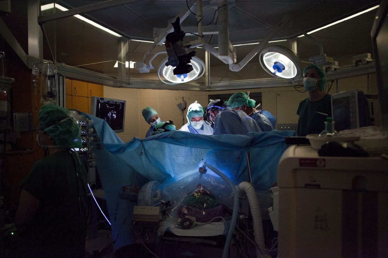 Directly at the table-a gynecologist surgeon, two neurosurgeons and operating sister Photo: Olga Pavlova
Spina Bifida is diagnosed by ultrasound at the twentieth week of pregnancy. It is generally accepted that the main cause of the disease is the lack of vitamins, especially folic acid. Even the non -specialist is clearly visible on the monitor of the tubercle, the bubble on the back of the fetus - the spine is, as it were, breaks down, the spinal cord and cerebrospinal fluid are noticeably bulging out. It is much more difficult to notice, although it often happens that the hernia, as it were, pulls the entire spinal cord along the entire length of the spine and, as it were, draws the occipital parts of the brain into the spine.
This is called Arnold-Kiary II syndrome. When children with Kiary syndrome are born, they experience a constant headache, the skull bursts from the inside, the head becomes so huge that there is no strength to tear it off the pillow. Usually they install a shunt in the head to take a liquid that bursts the skull. Kiari syndrome is an indication for a prenatal operation. Embrions with Spina Bifida, which do not have a kiari syndrome, do not operate intrauterine. They are operated on immediately after birth.
In Russia, only two intrauterine operations of Spina Bifida were carried out, and both, unfortunately, are unsuccessful: in the first case, the child died, in the second was born seriously paralyzed. The traditional method of surgical treatment Spina Bifida is to operate on the hernia immediately after the birth of the child. In Russia and third world countries, many neurosurgeons claim that there is no need to operate children with Spina Bifida. This opinion contradicts the results of objective research.
Children with Kiary syndrome experience a constant headache, the skull bursts from the inside, the head becomes so huge that there is no strength to tear it off the pillowThe fundamental here is the study of the MOMS (Management of Myelomeningocele Study), held in the United States from 2003 to 2010. The three largest clinics are the Philadelphia Children's Hospital, the University of Vanderbilt and the University of California at San Francisco divided patients with a diagnosis of “a defect in the formation of the fetal nervous tube” into two groups of 100 people each. The first group was done intrauterine operations, the second group - postnatal (that is, after birth). It turned out that if you operate the child with Spina Bifida postnatically, then in 98% of cases the child dies or needs to establish a shunt in the head. In the case of a fetal (that is, intrauterine) operations, mortality or the need to establish a shunt in the head decrease to 68%. In other words, if you operate the child with Spina Bifida and Kiary syndrome postnatally, then the child will almost certainly be a difficult disabled person. And if you operate in intrauterine, there are 32% of the chances of saving your head and avoid paralysis.
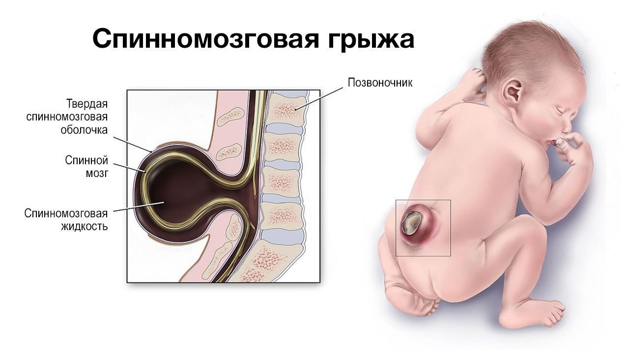 Spina Bifida Illustration: Centers for Disease Control and Prevention; Aksana Zinchenko
In addition, the development of motor functions in infants who have undergone intrauterine operations are evaluated much higher in the Beille scale than in babies operated on after birth.
On December 7, 2010, an independent committee that controlled the MOMS recommended the study to stop due to the fact that intrauterine operations are obviously more effective than postnatal. By the time MOMS stopping from 200 patients, which were supposed to be included in the study, 158 were operated on.
This is not a panacea, not a wonderful healing. Domestic operations are not shown to everyone. There are contraindications. Additional risks of uterine rupture or placenta exfoliation for a woman appear. But the intrauterine operation is a thirty percent chance for an unborn child to avoid disability, and for those who will not be able to avoid disability, this is a chance to reduce the degree of pathological changes.
The most blood supply organ
To do a prenatal operation, two anesthesia is used at once - general and epidural. Under general anesthesia, in fact, they operate. Epidural anesthesia is turned on when the operation is completed so that the uterus of the awakened patient does not decrease too much from postoperative pain, and the fetus does not die.
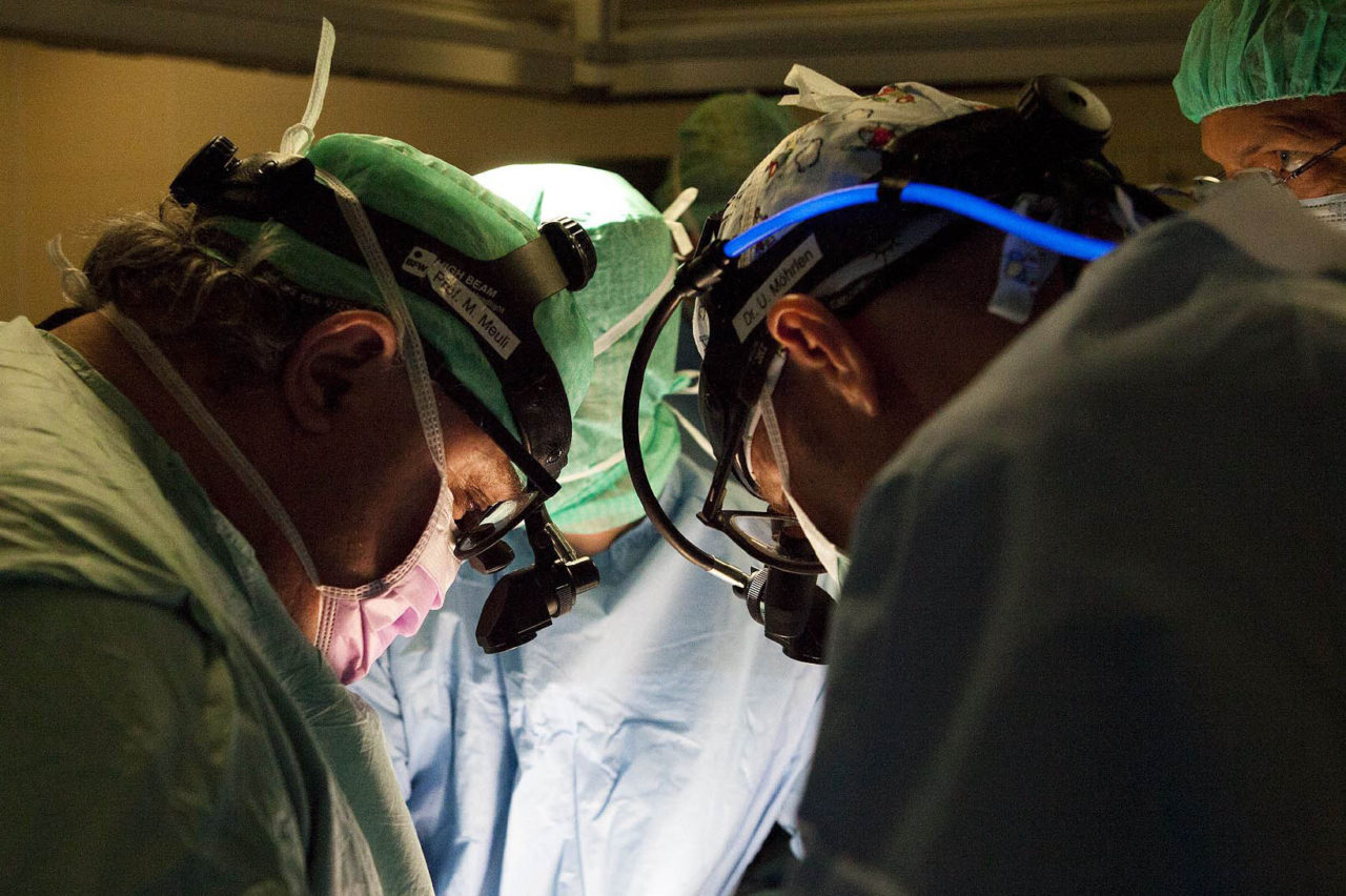 Professor Moil (on the left) says that even a good neurosurgeon can do intrauterine operations on his own only after he operated on the supervisions of a more experienced colleague photo: Olga Pavlova
Disord the peritoneum. The uterus almost get out. To get the uterus, a special extractor is used, something like a plastic bag or a toy basketball basket that covers the uterus from all sides and pulls up, and the ring rests on the outside on the stomach and does not allow the uterus to fail.
A pregnant uterus is the most blood supply organ in the human body after the brain. Therefore, you can’t just cut the uterus without receiving a blood fountain. In the uterus, a small hole is made with an electric knife, which simultaneously cuts and solders the vessels, and then a stapler is introduced into the incision. And before dissecting the uterus, the wall of the uterus and the amniotic fluid are sewn on both sides of the future section. The site’s place is determined using the ultrasound apparatus very carefully, so that in no case the incision does not affect the placenta and does not even pass near it.
The red liquid that splashes out when the uterus is dissected is amniotic fluid. The loss of amniotic fluid is not dangerous: as soon as the uterus is dissected, a tube is lowered into it, from which a Ringer's solution is constantly pushing - it replaces water, irrigates the fetus during the operation and remains in the uterus when the uterus is sewn.
When the uterus is dissected, and the edges of the cut are treated so that there is no bleeding, the gynecologist surgeon simply brings the fetus to the section and holds so that it is convenient to operate neurosurgeons.
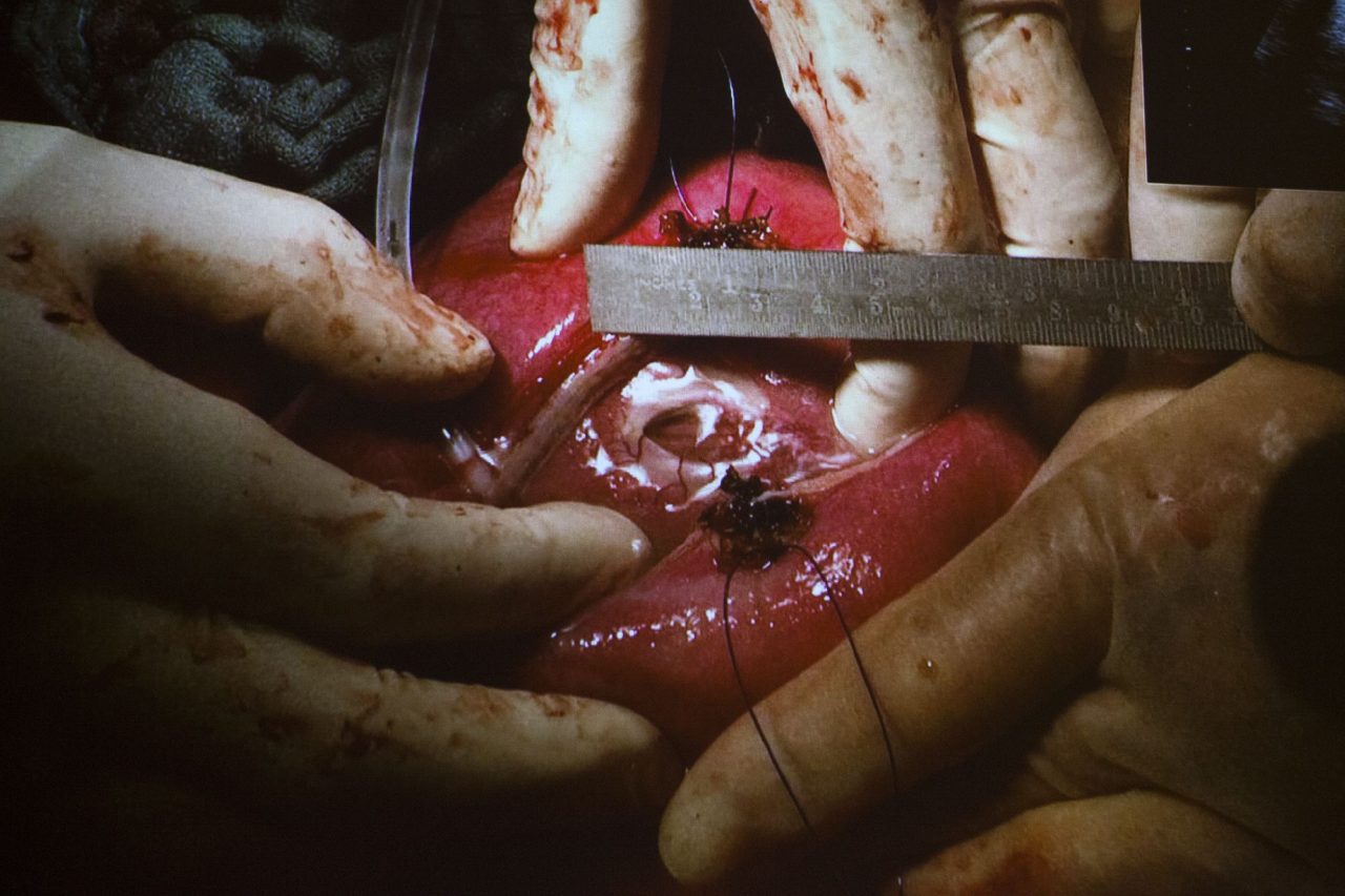 The gynecologist surgeon brings the fetus to the section in the uterus. This is what Spina Bifida looks like. Shortion from the monitor screen photo: Olga Pavlova
This is a very subtle operation. At the 24th week of pregnancy, the fruit is the size of a newborn kitten, and a hernia on his back is no more than a pea. The skin is dissected, the cerebrospinal fluid flows from it, and at that moment the naked eye shows how the spinal cord is drawn into the spine. And the ultrasound shows how the occipital parts of the brain are released and returned to their place inside the skull. Professor Moil still tried to show me how the spinal cord roots are being laid out in places, but it seems that a person is not able to see this without a special education.
At the 24th week of pregnancy, the fruit is the size of a newborn kitten, and a hernia on his back is no more than a peaWhen the spinal cord returns to its place inside the spine, the crevice above it is not fastened. Any attempt to fasten the split spine would lead to the impossibility of its further growth. Therefore, the spinal cord settled by the hernia is simply closed from the outside with the skin in the hope that the vertebrae that are not bursting will grow over by themselves. If the hernia is large, the skin on the back of the fetus often cannot be pulled and sewn enough. Then they use a piece of artificial epidermis, and after birth it will need to be replaced by the natural skin of the child, that is, the baby is waiting for another operation.
Now - sew the uterus. This is not an easy task. Pregnancy after the operation will continue for another three months, the uterus will stretch, the seams can be dispersed. Therefore, the edges of the cut are sewn, but the large area of the uterus around the cut is stitched with threads and then pulled together, as a duffel bag or bag for replaceable shoes is pulled.
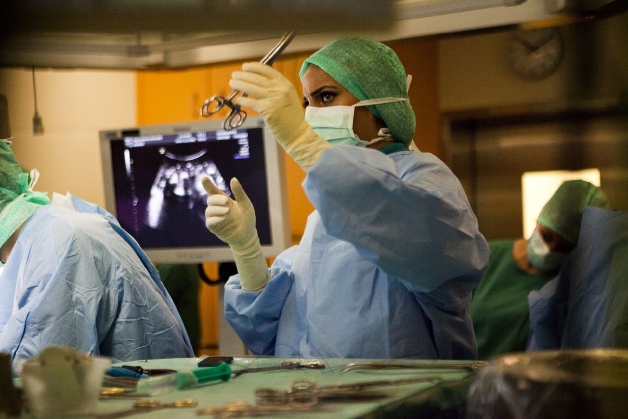 Each action is performed under the control of the ultrasound so that in no case do not touch the placenta photo: Olga Pavlova
The operation ends on this. If unexpected complications do not occur, it lasts two and a half to three hours. Professor Moil says that even a good neurosurgeon can do intrauterine operations on his own only after he operated on the supervision of a more experienced colleague at once. Each operation of the professor contains neurosurgeons and gynecologists from different cities of Europe. Moil willingly shares the methodology.
Take the child
About a day after the operation, when the patient has already woken up, epidural anesthesia continues. Another two days later you can get up and walk. But you can’t leave home. The intrauterine operation, which increases the child’s chances to be born more or less healthy, nevertheless increases the risks for a pregnant woman. They are afraid of uterine ruptures, placental exfoliation, premature birth. Therefore, the patients spend the remaining three months of pregnancy in a guest house, about 200 meters from the clinic. Between the 36th and 38th weeks, pregnancy is allowed by Caesarean section.
Usually a newborn child with Spina Bifida, even if the intrauterine operation was successful, differs from a healthy baby. He is, as it were, folded in half - legs to the head.
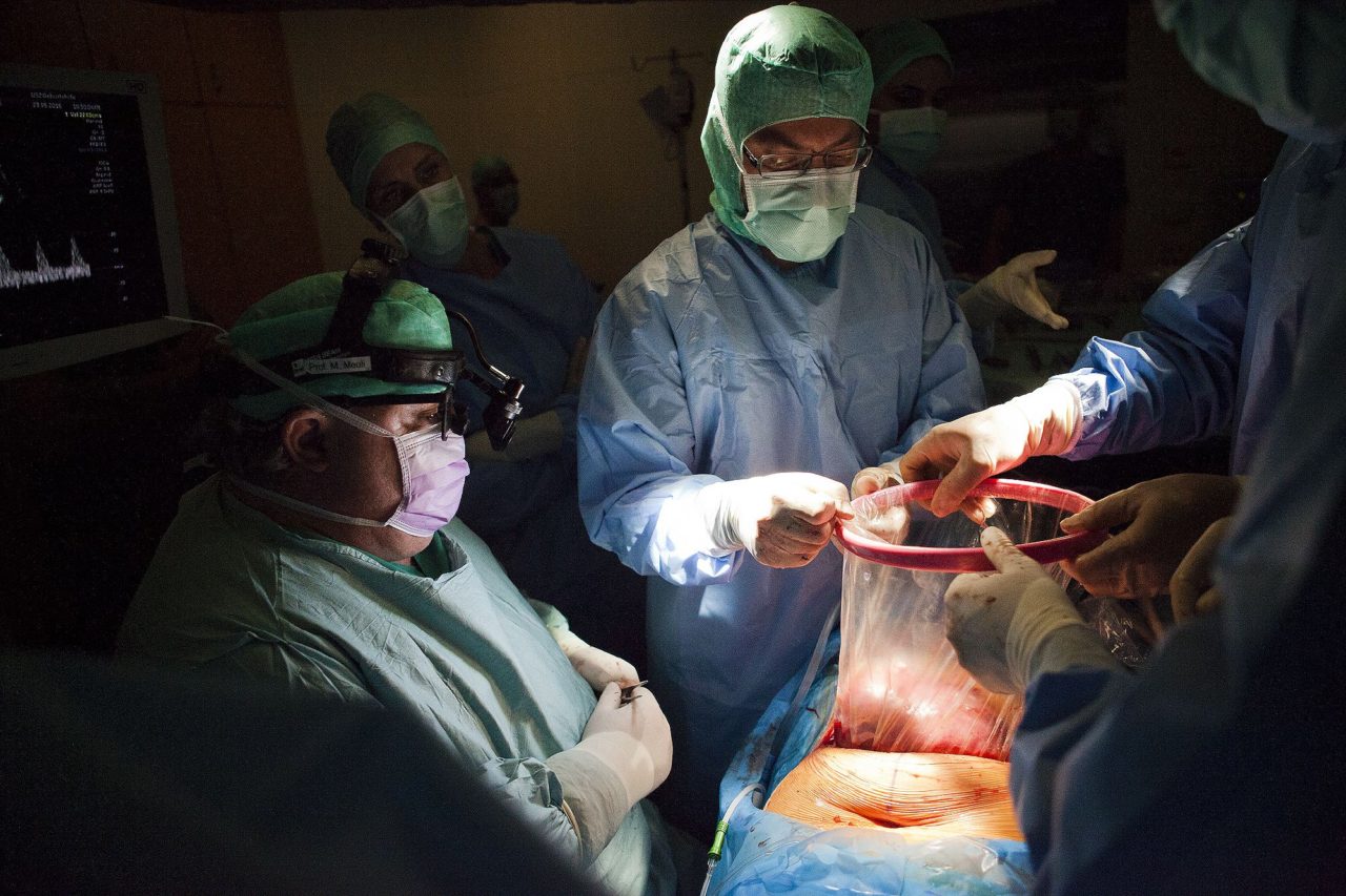 From the abdominal cavity, the uterus is taken out with an extractor similar to a plastic bag or a toy basketball basket photo: Olga Pavlova
The first person who, after cesarean section, brings the child to his mother is a rehabilitologist, a physical therapist. His task is to explain to his mother how to take a child in half, how to put a child who has an unprotected spinal cord right under his skin, how to feed a child who cannot be held under his back.
A newborn child with Spina Bifida, even if the operation was successful, differs from a healthy baby. He is, as it were, folded in half - legs to the headThe first week of life, baby and mother are carried out in the intensive care unit, that is, in intensive care. Here, for children with Spina Bifida, there are also special rules: for example, a blood test cannot be taken, like healthy children, from the heel, or you can - from their heads. Because it is not known about children with a hernia of the spinal cord how sensitive the newborn legs are, and whether the injection will lead to the heel, which does not feel anything, to serious hemorrhagic disorders.
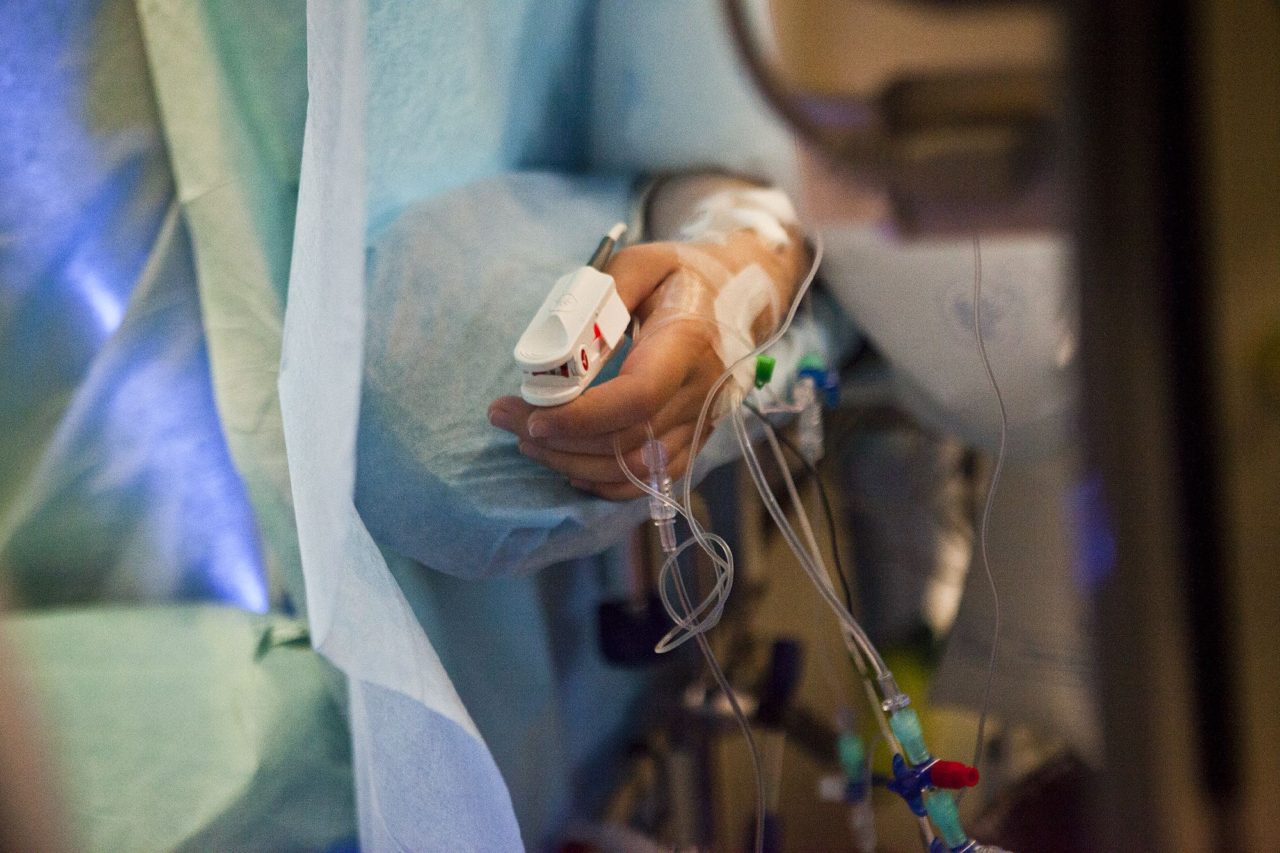 The patient receives general anesthesia during surgery and epidural immediately after the operation is completed by the photo: Olga Pavlova
Rehabilitation, physical therapy begins with the first day of life. The main meaning of rehabilitation is that, despite the surgery and vulnerability of the spinal cord, to provide the baby with all the activity that he has considered to be by age. Massage for children with Spina Bifida is not done. Never.
Ensuring the child activity is not an easy task. As a rule, children with Spina Bifida are sluggish and relaxed. In addition, even a successful operation can lead to complications, dermoid and epidermal cysts. And then additional operations will be required, and each of them at least excludes motor activity for several days.
The main thing, Professor Moil says, to understand that the intrauterine operation is not a one -time intervention that ensures recovery.
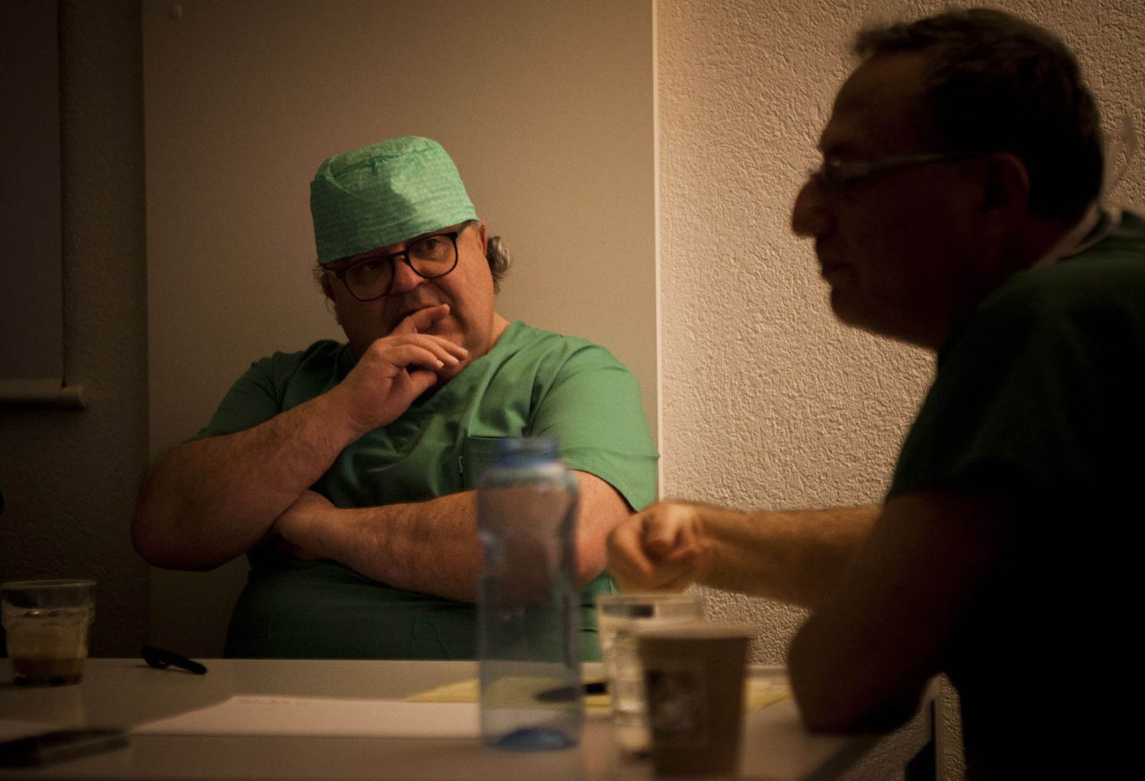 Professor Martin Miliy was the first to operate on the child with Spina Bifida in intrauterine and did 32 such operations. After each operation, the whole team gathers in a one and a half hour “flight analysis” photo: Olga Pavlova
Successfully conducted intrauterine operation is a very promising, but only the beginning of the treatment of a child with a hernia of the spinal cord. Practically, that a child with a hernia of the spinal cord grows more or less healthy, he needs several operations, rehabilitation and social adaptation throughout his life.
In the treatment and rehabilitation of children with Spina Bifida, a revolution has occurred, but only with charitable means to apply the achievements of this revolution in Russia. Therefore, we ask you to subscribe to regular donations in favor of the Spina Bifida program. The program will pay for operations to pregnant women and organize the rehabilitation of children with the consequences of the hernia of the spinal cord. Even a small amount of money, sacrificed by you regularly, can change the lives of thousands of children. There is a huge difference - to lie with a blurry skull or go to school, play with friends, live an active life.