
Translated material
Miniature organs
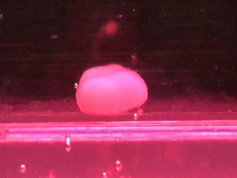
The technology for growing miniature human organs from stem cells began to actively develop only in the last decade. However, scientists have already been able to get analogues of the heart, kidneys, brain, stomach, lungs, retina, large and small intestines in laboratory conditions. They have groups of differentiated cells, similar to what are in full -sized organs.
To get an organoid, stem cells are placed on an environment that allows them to form a three -dimensional structure. There, they are self -organized and differentiated into cells of various types, repeating with some degree of accuracy the structure and even the functions of the real organ. Such organoids already serve to test drugs, but their role is no less important for fundamental studies, since with their help the genetic mechanisms of the formation of these organs during the development of the embryo can be established. We once talked about such studies in the essay "Organs from the Drive."
The development of any body is determined by a complex algorithm that provides for the inclusion and disconnection of specific genes at the right points. Scientists are just beginning to recognize the details of this program. Allows you to do this new technology-RNA sequencing from a single cage ( Single-Cell RNA Sequencing ). Reading RNA molecules makes it possible to determine which genes work at the moment, since it is with the help of the so -called information, or matrix RNAs that the information encoded in the genes is transmitted to ribosomes, where protein synthesis occurs. RNA is a short -living molecule, so a specific matrix RNA can be found only during the work of the gene related to it, not earlier and no later.
Therefore, scientists are grown from stem cells placed in the volumetric environment, the organoid and in the process of its development determine, securing the RNA of individual cells, what genes and how active are at the moment. Jason Spence development specialist from the University of Michigan says that sequencing of single cells is a great way to describe these processes with a sufficient degree of severity.
The use of organoids allows, moreover, much easier than, for example, research on laboratory animals, to apply various ways of influencing the genetic activity of cells. You can delete or insert individual genes using specially designed viruses or use the point editing method of CRISPR/CAS9 genome. And then look at what effect these changes caused. Biologists even learned to infect organoids with various bacterial or viral infections to determine the molecular mechanism of the disease. Now, for example, the effect on the brain of the ZIK viral fever is being studied . In addition, systems of joint cultivation of several organoids were developed, reproducing the structure of the body, including a network of neurons and cells of the immune system.
Last week, in the journal Nature published the most detailed study of the formation of a miniature liver from stem cells from stem cells. One of its authors - Takanori Takebe, working at the Universities of Iokogama and Tsincinnati - was interested in whether the artificially grown liver tissue can be used for transplantation to patients. He learned to successfully grow mini-organizations in his laboratory of only a few millimeters of pluripotent stem cells, which were differentiated into hepatocyte predecessions, mesenchymal and endothelial cells.
But he understood that the liver from the Petri cup could differ from the organ of natural origin. The attention of Takoeba was attracted by the work of Barbara Treutlein from the Institute of Molecular Cellular Biology and Genetics of the Society of Max Planck. Barbara leads the laboratory that specializes in sequencing of the RNA of single cells. In the work that Takebe drew attention to, she investigated the activity of genes in the formation of lungs in the embryos of bats. Takanori Takoeba invited her to jointly study the genetic mechanisms of growth of mini -rope from stem cells. Scientists were most interested in the interaction of different types of cells during the formation of the organ, because sometimes a protein isolated by a neighboring cell of a different type is a signal for the launch of any gene in the cell. Among the leading authors of the work were also Keisuke Sekine from Jokogama and J. Gray Camp from the Department of evolutionary genetics of the Institute of Evolutionary Anthropology of the Society of Max Planck.
According to the Takanori Takoeba method, miniature liver were grown, and at different stages of their development, researchers took cells and sequenced all RNA molecules from them, encoding proteins, determining the activity of the genes. Each time they received a complete set of active transcription factors (proteins that control the work of other genes), signal proteins and receptors involved at this particular moment.
For comparison, genes were also studied in the cells of human embryos and in the liver cells of an adult. According to the data obtained, the patterns of the work of genes in organoids are very close to the processes in the natural embryonic liver, but differ from the liver liver.
The liver organoid, grown by researchers from pluripotent human stem cells. Hepatocytes are painted with green, the cells of blood vessels are red.
In particular, for the first time in history, the authors managed to determine proteins that provide communication between different types of cells in a developing organoid. To test their results, the researchers created many new liver, but in their development, inhibitors that block the effect of signal proteins were added to the environment. This allowed scientists to turn off or turn on the processes of cell differentiation and organ formation of their own free will.
They also managed to establish the role of hypoxia - lack of oxygen - in the process of organoid growth. When the accumulation of cells becomes too large, those cells that are inside begin to experience oxygen deficiency. This forces cells that should give rise to blood vessels, begin the production of proteins responsible for this process. If after that, transplant the organoid into the liver of a laboratory mouse, it will be able to connect his forming vessels to its blood system.
“The possibility of creating a bioenger transplantable liver or liver tissue will be very useful for people suffering from liver diseases, for saving the lives of whose innovative methods of treatment are needed,” Takanori Takebe commented on the results obtained. “Our data give a new, detailed understanding of intercellular communication between developing liver cells and show that we can create fragments of the human liver, which are very surprisingly close to formations from embryonic cells that appear during the natural development of a person.”
In May of this year, Nature Cell Biology published another job , which checked the possibility of using miniature lungs grown in the laboratory to study viral respiratory studies and cystic fibrosis. The team of researchers from Colombian University was led by Professor Hans -Willem Snoeck. Scientists have grown model organoids from pluripotent stem cells, having achieved that analogues of branches of the bronchi, ending with alveoli, have analogues of them. Then the organoids exposed the virus or, editing the cellular genome, reproduced the mutation responsible for cystic fibrosis. In both cases, they observed the effects characteristic of this disease, which means that such minimal ones can be used in search of effective treatment methods.
Also this year, a group of scientists from the United States began to use miniords in the treatment of prostate cancer. Doctors under the leadership of Hatem Sabaawy from the Institute of Racian University Studies (Rutgers Cancer Institute of New Jersey) decided to grow model tumors from cells taken from patients and exposed to drugs proposed for the treatment of these patients. If the drug shows its effectiveness, it will be given to the patient.
Cultures of tumor cells for testing various treatment products have been grown for a long time, but researchers believe that flat tumor tissue in Petri's cup does not sufficiently reflect the complexity of the tumor and poorly predicts how patients will respond to treatment. Therefore, they decided to build three -dimensional analogues of the organ affected by the tumor. Researchers also intend to sequenate DNA of tumor tissue to create a bank of genetic profiles that can be used to treat other patients.
Professor Hans Clevers (Hans Clevers) from the Hububrecht Institute of the Netherlands Royal Academy of Sciences currently leads a similar project, which explores tumors of the colon. He says that although the study is at an early stage, the results obtained with the first patients look promising. According to Clever, laboratory studies allow you to choose the most effective drug for a particular patient and avoid the use of those drugs to which the cells of this tumor are stable. By the end of the year, two more projects for studying cancer on organoids will be started in the Netherlands, one will be dedicated to colorectal cancer, the other - cancer of the mammary glands.
Jatin Roper, head of the Center for Studies of the Hereditary Board of the Gastrointestinal Protection in the Medical Center for Tafts in Boston, combines the use of organoids with research on laboratory animals. Minor -organs that simulate the tissue of the large intestine with a tumor are grown in the laboratory, and then implanted into the intestines of the mouse. There, tumor cells interact with other intestinal cells, which allows researchers to observe cancer in a more natural environment, various genetic options are reproduced using CRISPR/CAS9 technology.