
Translated material
How to identify metastases
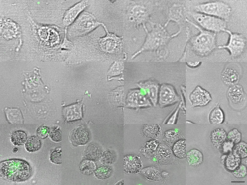
Scientists of the Moscow Physics and Technical Institute tested the effectiveness of a new method for assessing the risk of tumor metastasis. Having found out that mobile and plastic cancer cells have “increased appetite”, the researchers offered them fluorescent nanoparticles. It turned out that the more aggressive the tumor, the more particles it absorbs and the more intense the glow of the tissue sample under the microscope will be. This inexpensive and technically simple method will help to much quickly choose the optimal therapy for the patient for a particular stage of the disease. The study was spoke about the study by the press service of the MIPT.
Today, cancer is one of the main causes of mortality around the world. The culprit of more than 90 % of all deaths associated with cancer are metastases. Prediction of the development of metastases is a complex task: the main predictors of metastasis are the state of the lymph nodes, the size of the tumor and histological assessment. However, unfortunately, these predictors are not infallible.
The approach proposed by researchers from the laboratory of personalized chemo-ray MFTi therapy is based on the features of cancer cells found during previous studies: preparing to introduce healthy tissues, they start the process of reorganization of the actin cytoskeleton of the cell. Since the actin cytoskeleton also participates in the process of endocytosis - the absorption of the external cell material, the hypothesis was put forward that the metastasizing cancer cells increase the ability to capture particles of a certain size. Thus, tracking the amount of absorbed particles, cancer cells can be distinguished from healthy ones. And even to understand how far the process has gone, determining the percentage of these high -interastazing (well absorbing particles) as part of all tumor cells taken from the patient.
As “tags”, scientists have chosen nanoparticles modified by polystyrene carboxylate, loaded with fluorescent dyes. Such particles are widely available and have already been used to study the processes of endocytosis in cells, but without taking into account the connection with the risk of tumor metastasis. Under the influence of light with a certain wavelength, dyes glow, and the tags become clearly visible against the background of other cells and tissues. This property underlies the method of fluorescent microscopy. The sizes of particles were 100 and 200 nanometers, since smaller ones, firstly, are more expensive, and secondly, would require the use of more sensitive equipment.
The subjects were the cancer cells of the breast with a low and high risk of metastasis, as well as benign cells. Exploring them under a fluorescent microscope, scientists have found that benign cells do not absorb nanoparticles and do not even hold them on their cell wall. Cancer cells, on the contrary, hold the tags and absorb, and cells with a higher risk of metastasis do this more intensively.
Julia Merher, a senior researcher at the Laboratory of personalized chemo-ray MFTi therapy, says: “After analyzing the many images, we found out that larger particles (200 Nm) penetrate into cancer cells more efficiently than small ones that mainly remain on the cell wall. At the same time, an example of a more aggressive tumor absorbed almost twice as many particles than the cells of a benign tumor. ”
Based on the data obtained in the experiment, scientists recommend the use of particles with a size of 200 nm, since with their help you can distinguish not only metastatic and benign cells, but also cells with various metastatic potential. In the future (after additional studies), it will be possible to evaluate the risks of a tendency to metastasis cells of a particular patient.
The new method that allows you to get by a small number of cells in the patient, which is less traumatic and painful, inexpensive and fast: the answer can be obtained in just an hour, while traditional histological examination requires several weeks. It is hardly worth talking about how important this time is for the patient, because the sooner the treatment is started, the higher the chance of recovery.
The study is published in the journal Scientific Reports.